Bassett Collection of Stereoscopic Images of Human Anatomy
Dissection of posterior aspect of left thigh
Deep relations of sciatic nerve in popliteal fossa
Image #183-5
KEYWORDS: Muscles and tendons, Thigh, Vasculature.
Creative Commons
Stanford holds the copyright to the David L. Bassett anatomical images and has assigned Creative Commons license Attribution-Share Alike 4.0 International to all of the images.

For additional information regarding use and permissions, please contact the Medical History Center.
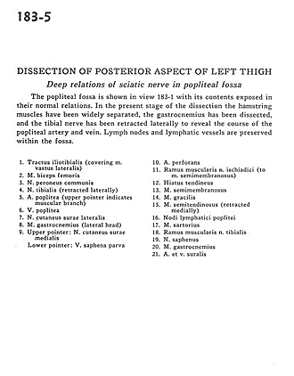
Dissection of posterior aspect of left thigh
Deep relations of sciatic nerve in popliteal fossa
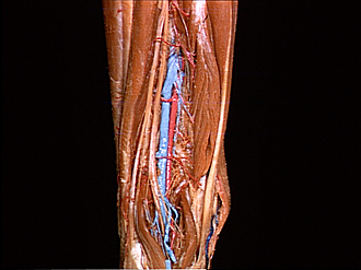
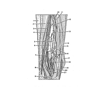
The popliteal fossa is shown in view 183-1 with its contents exposed in their normal relations. In the present stage of the dissection the hamstring muscles have been widely separated, the gastrocnemius has been dissected, and the tibial nerve has been retracted laterally to reveal the course of the popliteal artery and vein. Lymph nodes and lymphatic vessels are preserved within the fossa.
- Iliotibial tract (covering vastus lateralis muscle)
- Biceps femoris muscle
- Common peroneal nerve
- Tibial nerve (retracted laterally)
- Popliteal artery (upper pointer indicates muscular branch)
- Popliteal vein
- Lateral cutaneous sural nerve
- Gastrocnemius muscle (lateral head)
- Upper pointer: Medial cutaneous sural nerve Lower pointer: Lesser saphenous vein
- Perforating artery
- Muscular branch of sciatic nerve (to semimembranosus muscle)
- Adductor hiatus
- Semimembranosus muscle
- Gracilis muscle
- Semitendinosus muscle (retracted medially)
- Popliteal lymph node
- Sartorius muscle
- Muscular branch of tibial nerve
- Saphenous nerve
- Gastrocnemius muscle
- Sural artery and vein