Bassett Collection of Stereoscopic Images of Human Anatomy
Dissection of posterior aspect of left thigh
Relations of sciatic nerve in upper part of thigh
Image #183-3
KEYWORDS: Fascia, Peripheral nervous system, Thigh.
Creative Commons
Stanford holds the copyright to the David L. Bassett anatomical images and has assigned Creative Commons license Attribution-Share Alike 4.0 International to all of the images.

For additional information regarding use and permissions, please contact the Medical History Center.

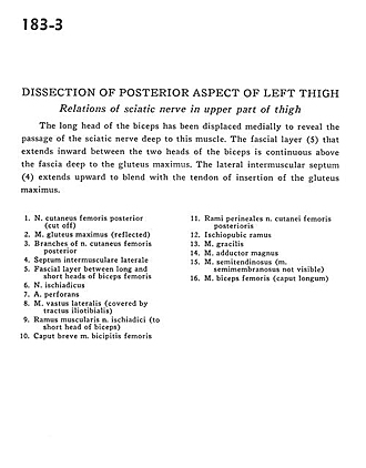
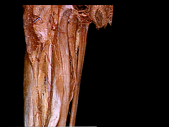
Dissection of posterior aspect of left thigh
Relations of sciatic nerve in upper part of thigh
The long head of the biceps has been displaced medially to reveal the passage of the sciatic nerve deep to this muscle. The fascial layer (5) that extends inward between the two heads of the biceps is continuous above the fascia deep to the gluteus maximus. The lateral intermuscular septum (4) extends upward to blend with the tendon of insertion of the gluteus maximus.
- Posterior femoral cutaneous nerve (cut off)
- Gluteus maximus muscle (reflected)
- Branches of posterior femoral cutaneous nerve
- Lateral intermuscular septum
- Fascial layer between long and short heads of biceps femoris
- Sciatic nerve
- Perforating artery
- Vastus lateralis muscle (covered by iliotibial tract)
- Muscular branch of sciatic nerve (to short head of biceps)
- Short head of biceps femoris muscle
- Perineal branch of posterior femoral cutaneous nerve
- Ischiopubic ramus
- Gracilis muscle
- Adductor magnus muscle
- Semitendinosus muscle (Semimembranosus muscle not visible)
- Biceps femoris muscle (long head)