Bassett Collection of Stereoscopic Images of Human Anatomy
Dissection of posterior aspect of left thigh
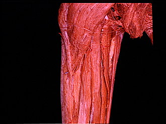
Posterior muscles of upper part of thigh, close-up view
Image #182-7
KEYWORDS: Muscles and tendons, Thigh.
Creative Commons
Stanford holds the copyright to the David L. Bassett anatomical images and has assigned Creative Commons license Attribution-Share Alike 4.0 International to all of the images.

For additional information regarding use and permissions, please contact the Medical History Center.


Dissection of posterior aspect of left thigh
Posterior muscles of upper part of thigh, close-up view
The specimen shown in the preceding photograph is illustrated here in a close-up view of the upper thigh.
- Ischial tuberosity
- Position of greater trochanter
- Gluteus maximus muscle
- Inferior cluneal nerve
- Posterior femoral cutaneous nerve (note cutaneous branches passing medially that penetrated fascia lata to reach skin)
- Fascial layer between long and short heads of biceps femoris (not lateral intermuscular septum)
- Iliotibial tract
- Biceps femoris muscle (upper pointer, long head; lower pointer, short head)
- Ischiorectal fossa
- Urogenital diaphragm
- Perineal branch of posterior femoral cutaneous nerve
- Gracilis muscle
- Adductor magnus muscle
- Semitendinosus muscle
- Semimembranosus muscle