Bassett Collection of Stereoscopic Images of Human Anatomy
Dissection of posterior aspect of left thigh
Superficial vessels; fascia lata; posterior femoral cutaneous nerve
Image #182-3
KEYWORDS: Fascia, Peripheral nervous system, Thigh, Vasculature.
Creative Commons
Stanford holds the copyright to the David L. Bassett anatomical images and has assigned Creative Commons license Attribution-Share Alike 4.0 International to all of the images.

For additional information regarding use and permissions, please contact the Medical History Center.
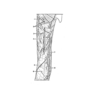
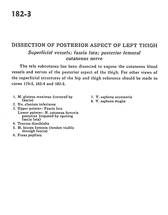
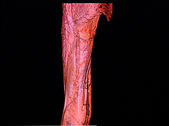
Dissection of posterior aspect of left thigh
Superficial vessels; fascia lata; posterior femoral cutaneous nerve
The tela subcutanea has been dissected to expose the cutaneous blood vessels and nerves of the posterior aspect of the thigh. For other views of the superficial structures of the hip and thigh reference should be made to views 179-5, 182-4, and 182-5.
- Gluteus maximus muscle (covered by fascia)
- Inferior cluneal nerve
- Upper pointer: Fascia lata Lower pointer: Posterior femoral cutaneous nerve (exposed by opening fascia lata)
- Iliotibial tract
- Biceps femoris muscle (tendon visible through fascia)
- Popliteal fossa
- Accessory saphenous vein
- Greater saphenous vein