Bassett Collection of Stereoscopic Images of Human Anatomy
Exploration of gluteal region and hip
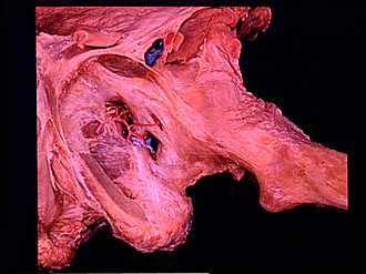
Capsule and ligaments of left hip joint, viewed from below
Image #181-6
KEYWORDS: Bones joints cartilage, Muscles and tendons, Thigh.
Creative Commons
Stanford holds the copyright to the David L. Bassett anatomical images and has assigned Creative Commons license Attribution-Share Alike 4.0 International to all of the images.

For additional information regarding use and permissions, please contact the Medical History Center.
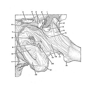

Exploration of gluteal region and hip
Capsule and ligaments of left hip joint, viewed from below
The femur has been abducted approximately 30
- Iliopsoas muscle (cut off)
- Femoral nerve
- Left pointer: Femoral sheath Right pointer: Femoral artery
- Inguinal ligament
- Spermatic cord
- Pectineus muscle (cut off)
- Body of pubis
- Obturator canal (note obturator nerve and artery emerging)
- Acetabular branch of obturator artery
- Obturator membrane
- Urogenital diaphragm
- Rectus femoris muscle (straight head)
- Iliopectineal bursa
- Iliofemoral ligament
- Hip articular capsule (part of capsule not well reinforced by ligaments)
- Lesser trochanter
- Tendon of insertion of obturator externus muscle
- Pubofemoral ligament
- Ischiofemoral ligament
- Left pointer: Acetabular notch Right pointer: Transverse acetabular ligament
- Ischial tuberosity
- Ischial ramus