Bassett Collection of Stereoscopic Images of Human Anatomy
Exploration of gluteal region and hip
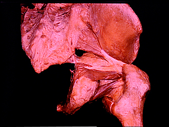
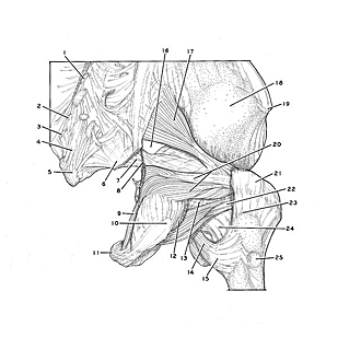
Piriformis and obturator muscles, posterior view of right side
Image #181-4
KEYWORDS: Bones joints cartilage, Muscles and tendons, Thigh.
Creative Commons
Stanford holds the copyright to the David L. Bassett anatomical images and has assigned Creative Commons license Attribution-Share Alike 4.0 International to all of the images.

For additional information regarding use and permissions, please contact the Medical History Center.

Exploration of gluteal region and hip
Piriformis and obturator muscles, posterior view of right side
This dissection has been photographed to illustrate the relations of the piriformis and the obturator internus and externus muscles to the capsule of the hip joint and to the bony pelvis and femur.
- Median sacral crest
- Sacral horn
- Coccygeal horn
- Superficial posterior sacrococcygeal ligament
- Coccyx bone
- Sacrotuberous ligament (cut off)
- Sacrospinous ligament
- Nerve to obturator internus muscle
- Obturator internus muscle
- Ischial tuberosity
- Body of pubis
- Quadratus femoris muscle (cut off)
- Hip articular capsule
- Iliopsoas muscle (tendon of insertion)
- Lesser trochanter
- Infrapiriform foramen
- Piriform muscle
- Ilium
- Tubercle of iliac crest
- Upper pointer: Tendon of obturator internus muscle Lower pointer: Inferior gemellus muscle (note absence of a superior gemellus muscle)
- Greater trochanter
- Tendon of insertion of obturator externus muscle
- Quadrate tubercle
- Neck of femur
- Trochanter tertius