Bassett Collection of Stereoscopic Images of Human Anatomy
Exploration of gluteal region and hip
Nerve to quadratus femoris muscle
Image #180-6
KEYWORDS: Muscles and tendons, Peripheral nervous system.
Creative Commons
Stanford holds the copyright to the David L. Bassett anatomical images and has assigned Creative Commons license Attribution-Share Alike 4.0 International to all of the images.

For additional information regarding use and permissions, please contact the Medical History Center.
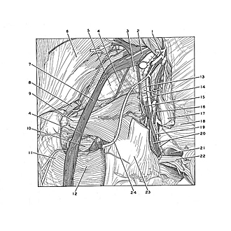
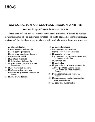
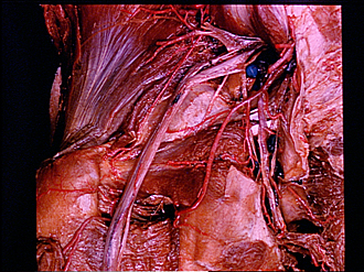
Exploration of gluteal region and hip
Nerve to quadratus femoris muscle
Branches of the sacral plexus have been elevated in order to demonstrate the nerve to the quadratus femoris (4) in its course across the posterior surface of the ischium deep to the gemelli and obturator internus muscles.
- Inferior gluteal artery
- Sacral plexus (elevated)
- Parietal pelvic fascia
- Nerve to Quadratus femoris muscle
- Body of ischium
- Gluteus minimus muscle
- Sciatic nerve (elevated)
- Piriform muscle (cut off close to insertion)
- Obturator internus muscle
- Obturator externus muscle
- Location of quadrate tubercle of femur
- Quadratus femoris muscle
- Internal pudendal artery
- Sacrospinous ligament
- Nerve to obturator internus
- Inferior rectal artery
- Sacrotuberous ligament (cut and reflected medially)
- Levator ani muscle
- Pudendal nerve
- Upper pointer: Pudendal canal Lower pointer: Lunate fascia (reflected to open pudendal canal)
- Ischiorectal fossa (anterior recess)
- Deep transverse perineal muscle
- Ischial tuberosity
- Artery supplying sciatic nerve