Bassett Collection of Stereoscopic Images of Human Anatomy
Exploration of gluteal region and hip
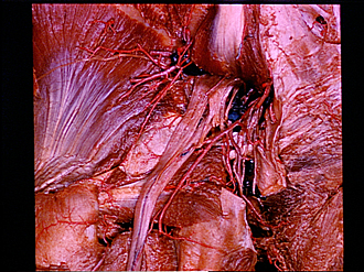
Contents of greater sciatic foramen
Image #180-5
KEYWORDS: Muscles and tendons.
Creative Commons
Stanford holds the copyright to the David L. Bassett anatomical images and has assigned Creative Commons license Attribution-Share Alike 4.0 International to all of the images.

For additional information regarding use and permissions, please contact the Medical History Center.


Exploration of gluteal region and hip
Contents of greater sciatic foramen
The tendon of the piriformis (9) has been divided near its insertion to allow the muscle (1) to be reflected upward. The sacrotuberous ligament (24) has also been divided and its upper part reflected medially. The sacral plexus is partially exposed.
- Piriform muscle
- Sacral plexus
- Superior gluteal artery and vein
- Right pointer: Inferior gluteal nerve Left pointer: Posterior femoral cutaneous nerve
- Superior gluteal nerve
- Gluteus minimus muscle
- Body of ischium (covered by periosteum)
- Sciatic nerve
- Piriform muscle (tendon of insertion, cut off)
- Greater trochanter (covered by tendon of Gluteus medius muscle)
- Obturator internus muscle (tendon of insertion)
- Upper pointer: Obturator externus muscle Lower pointer: Medial femoral circumflex artery
- Sacral bone
- Inferior gluteal artery
- Inferior cluneal nerve (medial)
- Internal pudendal vein (cut off)
- Lymph node
- Perforating cutaneous nerve
- Nerve to obturator internus
- Inferior rectal nerve
- Upper pointer: Sacrospinal ligament Lower pointer: Pudendal nerve (slightly elevated)
- Internal pudendal artery and vein
- Levator ani muscle
- Sacrotuberous ligament (divided and retracted)
- Deep transverse perineal muscle
- Ischial tuberosity
- Gemelli muscles
- Artery supplying sciatic nerve
- Quadratus femoris muscle