
Bassett Collection of Stereoscopic Images of Human Anatomy
Exploration of the brain from the medial aspect
Retrolenticular and sublenticular parts of internal capsule; striate arteries
Image #18-4
KEYWORDS: Brain, Telencephalon, Vasculature.
Creative Commons
Stanford holds the copyright to the David L. Bassett anatomical images and has assigned Creative Commons license Attribution-Share Alike 4.0 International to all of the images.

For additional information regarding use and permissions, please contact the Medical History Center.
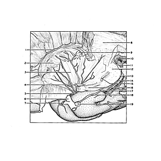
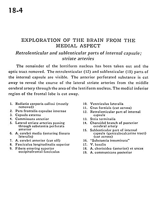
Exploration of the brain from the medial aspect
Retrolenticular and sublenticular parts of internal capsule; striate arteries
The remainder of the lentiform nucleus has been taken out and the optic tract removed. The retrolenticular (12) and sublenticular (15) parts of the internal capsule are visible. The anterior perforated substance is cut away to reveal the course of the lateral striate arteries from the middle cerebral artery through the area of the lentiform nucleus. The medial inferior region of the frontal lobe is cut away.
- Radiation corpus callosum (mostly removed)
- Frontal part internal capsule
- External capsule
- Anterior commissure
- Lateral striate arteries passing through anterior perforated substance
- Middle cerebral artery (entering lateral fissure)
- Anterior cerebral artery (cut off)
- Superior longitudinal fasciculus
- Fibers entering superior occipitofrontal fasciculus
- Lateral ventricle
- Fornix (crus) (cut across)
- Retrolenticular part of internal capsule
- Stria terminalis
- Choroidal branch of posterior cerebral artery
- Sublenticular part of internal capsule (geniculocalcarine tract) (cut across)
- "Substantia innominata"
- Basal vein
- Choroidal artery (anterior) and uncus
- Posterior communicating artery