Bassett Collection of Stereoscopic Images of Human Anatomy
Exploration of the brain from the medial aspect
Lateral geniculate body and geniculocalcarine tract
Image #18-1
KEYWORDS: Brain, Diencephalon.
Creative Commons
Stanford holds the copyright to the David L. Bassett anatomical images and has assigned Creative Commons license Attribution-Share Alike 4.0 International to all of the images.

For additional information regarding use and permissions, please contact the Medical History Center.

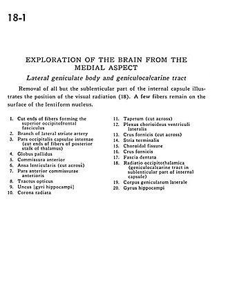
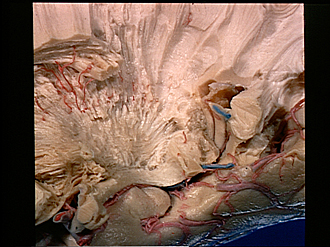
Exploration of the brain from the medial aspect
Lateral geniculate body and geniculocalcarine tract
Removal of all but the sublenticular part of the internal capsule illustrates the position of the visual radiation (18). A few fibers remain on the surface of the lentiform nucleus.
- Cut ends of fibers forming the superior occipitofrontal fasciculus
- Branch of lateral striate artery
- Occipital part internal capsule (cut ends of fibers of posterior stalk of thalamus)
- Globus pallidus
- Anterior commissure
- Ansa lenticularis (cut across)
- Anterior part of anterior commissure
- Optic tract
- Uncus (hippocampal gyrus)
- Corona radiata
- Tapetum (cut across)
- Choroid plexus lateral ventricle
- Fornix (crus) (cut across)
- Stria terminalis
- Choroidal fissure
- Fornix (crus)
- Dentate fascia
- Optic radiation (geniculocalcarine tract in sublenticular part of internal capsule)
- Lateral geniculate body
- Hippocampal gyrus