Bassett Collection of Stereoscopic Images of Human Anatomy
Exploration of gluteal region and hip
Gluteus maximus in relation to lumbosacral spine, posterior view
Image #179-6
KEYWORDS: Muscles and tendons.
Creative Commons
Stanford holds the copyright to the David L. Bassett anatomical images and has assigned Creative Commons license Attribution-Share Alike 4.0 International to all of the images.

For additional information regarding use and permissions, please contact the Medical History Center.
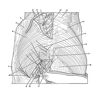
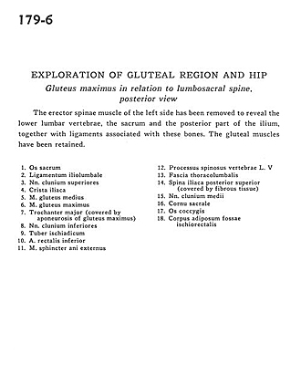
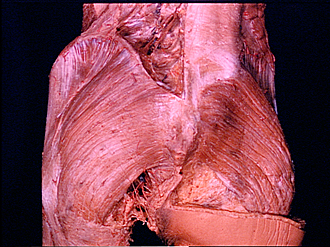
Exploration of gluteal region and hip
Gluteus maximus in relation to lumbosacral spine, posterior view
The erector spinae muscle of the left side has been removed to reveal the lower lumbar vertebrae, the sacrum and the posterior part of the ilium, together with ligaments associated with these bones. The gluteal muscles have been retained.
- Sacral bone
- Iliolumbar ligament
- Superior cluneal nerve
- Iliac crest
- Gluteus medius muscle
- Gluteus maximus muscle
- Greater trochanter (covered by aponeurosis of gluteus maximus)
- Inferior cluneal nerve
- Ischial tuberosity
- Inferior rectal artery
- External anal sphincter muscle
- Vertebral spinous process of L. V
- Thoracolumbar fascia
- Posterior superior iliac spine (covered by fibrous tissue)
- Medial cluneal nerve
- Sacral horn
- Coccyx bone
- Adipose body of ischiorectal fossa