
Bassett Collection of Stereoscopic Images of Human Anatomy
Exploration of gluteal region and hip
Superficial vessels and nerves of gluteal region
Image #179-5
KEYWORDS: Fascia, Peripheral nervous system, Vasculature.
Creative Commons
Stanford holds the copyright to the David L. Bassett anatomical images and has assigned Creative Commons license Attribution-Share Alike 4.0 International to all of the images.

For additional information regarding use and permissions, please contact the Medical History Center.

Exploration of gluteal region and hip
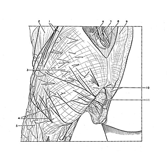
Superficial vessels and nerves of gluteal region
The tela subcutanea has been dissected to expose the numerous small arteries and veins that perforate the fascia lata to ramify superficially over the buttock. The superior (1) and inferior (4) clunial nerves have been dissected. The more laterally placed inferior clunial nerves (4, lower pointer) are derived from the posterior femoral cutaneous nerve, whereas those emerging more medially arise from the perforating cutaneous nerve (S2, S3). Middle clunial nerves are not clearly shown in this preparation but are demonstrated in the following view.
- Superior cluneal nerve
- Gluteus medius muscle
- Gluteus maximus muscle (covered with fascia lata)
- Inferior cluneal nerve
- Iliotibial tract
- Erector spinae muscle
- Sacral bone
- Dura mater (exposed within sacral canal)
- Thoracolumbar fascia
- Upper pointer: Anococcygeal nerve, Lower pointer: Coccyx bone (terminal part)
- Ischiorectal fossa