
Bassett Collection of Stereoscopic Images of Human Anatomy
Radiography
Left knee, posteroanterior view illustrating intercondylar fossa
Image #178-4
KEYWORDS: Bones joints cartilage, Knee, Thigh.
Creative Commons
Stanford holds the copyright to the David L. Bassett anatomical images and has assigned Creative Commons license Attribution-Share Alike 4.0 International to all of the images.

For additional information regarding use and permissions, please contact the Medical History Center.

Radiography
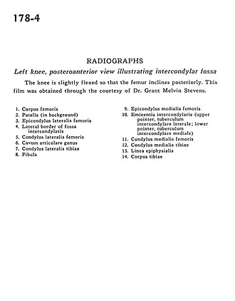
Left knee, posteroanterior view illustrating intercondylar fossa
The knee is slightly flexed so that the femur inclines posteriorly. This film was obtained through the courtesy of Dr. Melvin Grant Stevens.
- Body of femur
- Patella (in background)
- Lateral epicondyle of femur
- Lateral border of intercondylar fossa
- Lateral condyle of femur
- Knee articular space
- Lateral condyle of tibia
- Fibula
- Medial epicondyle of femur
- Intercondylar eminence (upper pointer, lateral intercondylar tubercle; lower pointer, medial intercondylar tubercle)
- Medial condyle of femur
- Medial condyle of tibia
- Epiphysial line
- Body of tibia