Bassett Collection of Stereoscopic Images of Human Anatomy
Osteology
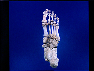
Articulated bones of left foot, inferior aspect
Image #177-6
KEYWORDS: Bones joints cartilage, Foot and toes.
Creative Commons
Stanford holds the copyright to the David L. Bassett anatomical images and has assigned Creative Commons license Attribution-Share Alike 4.0 International to all of the images.

For additional information regarding use and permissions, please contact the Medical History Center.

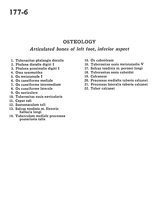
Osteology
Articulated bones of left foot, inferior aspect
- Tuberosity of distal phalanx
- Distal phalanx
- Proximal phalanx
- Sesamoid bone
- Metatarsal bone
- Medial cuneiform bone
- Intermediate cuneiform bone
- Lateral cuneiform bone
- Navicular bone
- Tuberosity of navicular bone
- Head of talus
- Sustentaculum tali
- Groove for flexor hallucis longus tendon
- Medial tubercle of posterior process of talus
- Cuboid bone
- Tuberosity of 5th metatarsal bone
- Groove for peroneus longus tendon
- Tuberosity of cuboid bone
- Calcaneus
- Medial process of tuberosity of calcaneus
- Lateral tubercle process of calcaneus bone
- Tuberosity of calcaneus