
Bassett Collection of Stereoscopic Images of Human Anatomy
Osteology
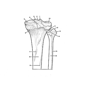
Right tibia and fibula, posterior view of upper parts
Image #176-7
KEYWORDS: Bones joints cartilage, Leg.
Creative Commons
Stanford holds the copyright to the David L. Bassett anatomical images and has assigned Creative Commons license Attribution-Share Alike 4.0 International to all of the images.

For additional information regarding use and permissions, please contact the Medical History Center.
Osteology
Right tibia and fibula, posterior view of upper parts
- Lateral intercondylar tubercle
- Intercondylar eminence
- Medial intercondylar tubercle
- Posterior intercondylar area
- Medial condyle of tibia (pointer indicates superior articular surface)
- Epiphysial line
- Soleal line
- Posterior surface
- Nutrient foramen
- Lateral condyle of tibia (pointer on superior articular surface)
- Articular surface of fibula
- Apex of fibula
- Head of fibula
- Articular surface of head of fibula
- Body of fibula
- Posterior surface