
Bassett Collection of Stereoscopic Images of Human Anatomy
Osteology
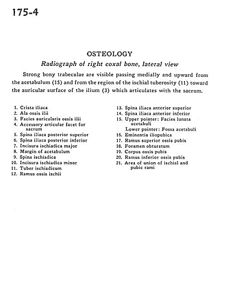
Radiograph of right coxal bone, lateral view
Image #175-4
KEYWORDS: Bones joints cartilage.
Creative Commons
Stanford holds the copyright to the David L. Bassett anatomical images and has assigned Creative Commons license Attribution-Share Alike 4.0 International to all of the images.

For additional information regarding use and permissions, please contact the Medical History Center.

Osteology
Radiograph of right coxal bone, lateral view
Strong bony trabeculae are visible passing medially and upward from the acetabulum (15) and from the region of the ischial tuberosity (11) toward the auricular surface of the ilium (3) which articulates with the sacrum.
- Iliac crest
- Iliac fossa
- Auricular surface for sacrum
- Accessory articular surface for sacrum
- Posterior superior iliac spine
- Posterior inferior iliac spine
- Greater sciatic notch
- Acetabular margin
- Ischial spine
- Lesser sciatic notch
- Ischial tuberosity
- Ischial ramus
- Anterior superior iliac spine
- Anterior inferior iliac spine
- Upper pointer: Acetabular lunate surface Lower pointer: Acetabular fossa
- Iliopubic (pectineal) eminence
- Superior pubic ramus
- Obturator foramen
- Body of pubis
- Inferior pubic ramus
- Area of union of ischial and pubic rami