
Bassett Collection of Stereoscopic Images of Human Anatomy
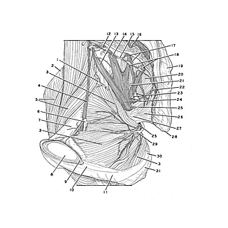
Dissection of male pelvis from a lateral approach
Obturator internus muscle and nerve supply, medial view
Image #172-7
KEYWORDS: Muscles and tendons, Peripheral nervous system.
Creative Commons
Stanford holds the copyright to the David L. Bassett anatomical images and has assigned Creative Commons license Attribution-Share Alike 4.0 International to all of the images.

For additional information regarding use and permissions, please contact the Medical History Center.

Dissection of male pelvis from a lateral approach
Obturator internus muscle and nerve supply, medial view
The pelvic diaphragm has been removed from the specimen and the obturator fascia has been stripped away.
- Iliac branch of iliolumbar artery
- Iliac crest
- Obturator internus muscle
- Obturator nerve
- Iliopsoas muscle
- Obturator artery
- Superior pubic ramus
- Pubic symphysis (sectioned)
- Pubic arcuate ligament
- Inferior pubic ramus
- Ramus of ischium
- Ramus communicans (to sacral nerve I)
- Lumbosacral trunk
- Sacral nerve I (ventral ramus)
- Sympathetic trunk (pointer on ganglion)
- Anterior (pelvic) sacral foramen (pointer indicates opening within sacral canal exposed by cutting through sacrum)
- Rami communicantes
- Left pointer: Sacral nerve II Right pointer: Sacral nerve III
- Sacrum
- Piriform muscle
- Inferior gluteal artery
- Sacral nerve IV
- Upper pointer: Inferior rectal nerve Lower pointer: Pudendal nerve
- Upper pointer: Branch of sacral nerve III to posterior femoral cutaneous nerve Lower pointer: Sciatic nerve
- Nerve to obturator internus muscle (indicated in two places)
- Coccygeus muscle
- Location of ischial spine
- Coccyx
- Internal pudendal artery
- Sacrotuberous ligament (cut off at attachment)
- Ischial tuberosity