Bassett Collection of Stereoscopic Images of Human Anatomy
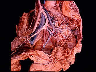
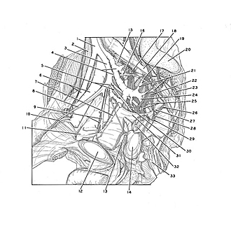
Dissection of male pelvis from a lateral approach
Blood vessels and nerves of right lateral pelvic wall
Image #172-4
KEYWORDS: Peripheral nervous system, Vasculature.
Creative Commons
Stanford holds the copyright to the David L. Bassett anatomical images and has assigned Creative Commons license Attribution-Share Alike 4.0 International to all of the images.

For additional information regarding use and permissions, please contact the Medical History Center.
Dissection of male pelvis from a lateral approach
Blood vessels and nerves of right lateral pelvic wall
Visceral branches of vessels and nerves have been cut away. The prostate and seminal vesicles have been retained in situ. The view is from the left and slightly anterior.
- Common iliac artery
- Psoas major muscle
- Internal iliac artery
- Upper pointer: Internal iliac vein (pointer at origin of superior gluteal vein) Lower pointer: Superior gluteal artery
- Upper pointer: Inferior gluteal artery Lower pointer: Internal pudendal artery
- External iliac artery and vein
- Obturator vein
- Lateral umbilical ligament
- Upper pointer: Obturator artery Lower pointer: Obturator nerve
- Inferior epigastric artery
- Superior pubic ramus
- Pubic symphysis (sectioned)
- Internal urethral opening
- Prostate
- Spinal branch iliolumbar artery
- Lateral sacral artery
- Sacral nerve I (ventral ramus)
- Pelvic surface of sacrum
- Right sympathetic trunk
- Articular surface of sacrum
- Lateral sacral veins
- Sacral nerve II (ventral ramus)
- Sacral nerve III
- Inferior gluteal vein
- Upper pointer: Pelvic splanchnic nerve (cut off) Lower pointer: Pudendal nerve
- Sacral nerve IV (ventral ramus)
- Fascia of piriform muscle (mostly removed)
- Pelvic splanchnic nerve (cut off)
- Nerve to Levator ani muscle
- Sciatic nerve
- Superior fascia of pelvic diaphragm (at posterior border of pelvic diaphragm)
- Seminal vesicle
- Anal canal