Bassett Collection of Stereoscopic Images of Human Anatomy
Dissection of male pelvis from a lateral approach
Sigmoid colon, rectum and anal canal opened in situ, left anterolateral view
Image #171-6
KEYWORDS: Anal canal, Large intestine.
Creative Commons
Stanford holds the copyright to the David L. Bassett anatomical images and has assigned Creative Commons license Attribution-Share Alike 4.0 International to all of the images.
For additional information regarding use and permissions, please contact the Medical History Center.
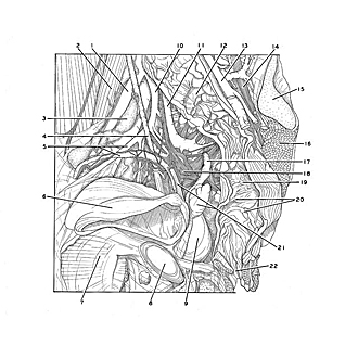
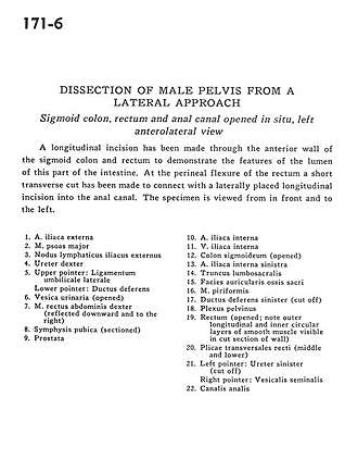
Dissection of male pelvis from a lateral approach
Sigmoid colon, rectum and anal canal opened in situ, left anterolateral view
A longitudinal incision has been made through the anterior wall of the sigmoid colon and rectum to demonstrate the features of the lumen of this part of the intestine. At the perineal flexure of the rectum a short transverse cut has been made to connect with a laterally placed longitudinal incision into the anal canal. The specimen is viewed from in front and to the left.
- External iliac artery
- Psoas major muscle
- External iliac lymph node
- Ureter right
- Upper pointer: Lateral umbilical ligament Lower pointer: Ductus deferens
- Urinary bladder (opened)
- Rectus abdominis muscle right (reflected downward and to the right)
- Pubic symphysis (sectioned)
- Prostate
- Internal iliac artery
- Internal iliac vein
- Sigmoid colon (opened)
- Left internal iliac artery
- Lumbosacral trunk
- Articular surface of sacrum
- Piriform muscle
- Ductus deferens left (cut off)
- Pelvic plexus
- Rectum (opened note outer longitudinal and inner circular layers of smooth muscle visible in cut section of wall)
- Plicae transversales of rectum (middle and lower)
- Left pointer: Ureter left (cut off) Right pointer: Seminal vesicle
- Anal canal


