Bassett Collection of Stereoscopic Images of Human Anatomy
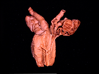
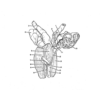
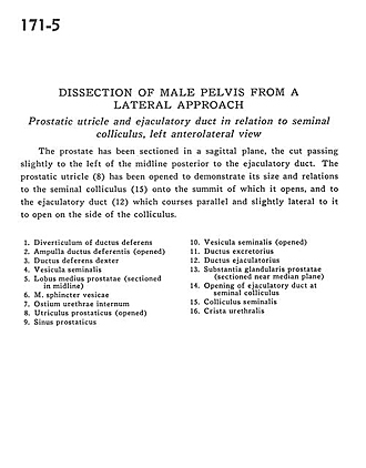
Dissection of male pelvis from a lateral approach
Prostatic utricle and ejaculatory duct in relation to seminal colliculus, left anterolateral view
Image #171-5
KEYWORDS:
Creative Commons
Stanford holds the copyright to the David L. Bassett anatomical images and has assigned Creative Commons license Attribution-Share Alike 4.0 International to all of the images.

For additional information regarding use and permissions, please contact the Medical History Center.
Dissection of male pelvis from a lateral approach
Prostatic utricle and ejaculatory duct in relation to seminal colliculus, left anterolateral view
The prostate has been sectioned in a sagittal plane, the cut passing slightly to the left of the midline posterior to the ejaculatory duct. The prostatic utricle (8) has been opened to demonstrate its size and relations to the seminal colliculus (15) onto the summit of which it opens, and to the ejaculatory duct (12) which courses parallel and slightly lateral to it to open on the side of the colliculus.
- Diverticulum of ductus deferens
- Ampulla of ductus deferens (opened)
- Ductus deferens right
- Seminal vesicle
- Middle lobe of prostate (sectioned in midline)
- Vesical sphincter muscle
- Internal urethral opening
- Prostatic utricle (opened)
- Prostatic sinus
- Seminal vesicle (opened)
- Excretory duct
- Ejaculatory duct
- Glandular substance of prostate (sectioned near median plane)
- Opening of ejaculatory duct at seminal colliculus
- Seminal colliculus
- Urethral crest