Bassett Collection of Stereoscopic Images of Human Anatomy
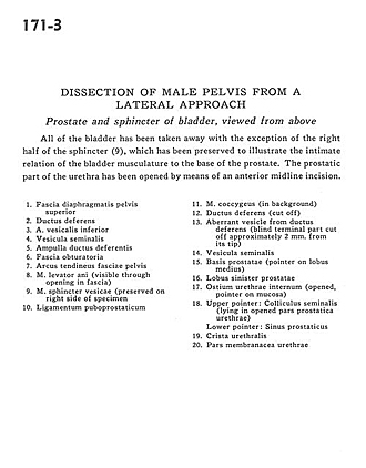
Dissection of male pelvis from a lateral approach
Prostate and sphincter of bladder, viewed from above
Image #171-3
KEYWORDS: Muscles and tendons, Urinary tract.
Creative Commons
Stanford holds the copyright to the David L. Bassett anatomical images and has assigned Creative Commons license Attribution-Share Alike 4.0 International to all of the images.

For additional information regarding use and permissions, please contact the Medical History Center.

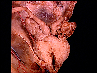
Dissection of male pelvis from a lateral approach
Prostate and sphincter of bladder, viewed from above
All of the bladder has been taken away with the exception of the right half of the sphincter (9), which has been preserved to illustrate the intimate relation of the bladder musculature to the base of the prostate. The prostatic part of the urethra has been opened by means of an anterior midline incision.
- Superior fascia of pelvic diaphragm
- Ductus deferens
- Inferior vesical artery
- Seminal vesicle
- Ampulla of ductus deferens
- Obturator fascia
- Tendinous arch of pelvic fascia
- Levator ani muscle (visible through opening in fascia)
- Vesical sphincter muscle (preserved on right side of specimen)
- Puboprostatic ligament
- Coccygeus muscle (in background)
- Ductus deferens (cut off)
- Aberrant vesicle from ductus deferens (blind terminal part cut off approximately 2 mm. from its tip)
- Seminal vesicle
- Base of prostate (pointer on middle lobe)
- Left lobe of prostate
- Internal urethral opening (opened, pointer on mucosa)
- Upper pointer: Seminal colliculus (lying in opened prostatic part of urethra) Lower pointer: Prostatic sinus
- Urethral crest
- Membranous part of urethra