Bassett Collection of Stereoscopic Images of Human Anatomy
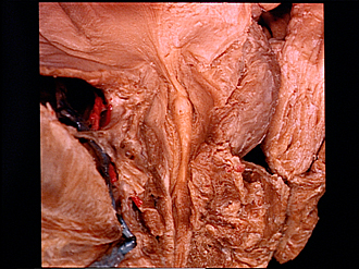
Dissection of male pelvis from a lateral approach
Interior of prostatic urethra; seminal colliculus, anterior view
Image #171-2
KEYWORDS:
Creative Commons
Stanford holds the copyright to the David L. Bassett anatomical images and has assigned Creative Commons license Attribution-Share Alike 4.0 International to all of the images.

For additional information regarding use and permissions, please contact the Medical History Center.


Dissection of male pelvis from a lateral approach
Interior of prostatic urethra; seminal colliculus, anterior view
A midline incision has been made beginning at the internal urethral orifice and extending downward through the isthmus of the prostate to open the prostatic and membranous parts of the urethra. The left lobe of the prostate has been pulled aside to provide adequate exposure of the urethral lumen.
- Pubovesicalis muscle (also see view 170-7)
- Vesical sphincter muscle
- Uvula of bladder
- Internal urethral opening
- Puboprostatic ligament (pubovesical part)
- Prostate (sectioned in midline)
- Puboprostatic ligament
- Sphincter muscle of urethra (cut ends separated, posterior part elevated)
- Interpubic disc
- Dorsal vein of the penis
- Rectum
- Left lobe of prostate
- Isthmus of prostate (sectioned)
- Openings of ejaculatory ducts (opening on seminal colliculus)
- Prostatic utricle (opening on summit of seminal colliculus)
- Prostatic part of urethra (pointer indicates prostatic sinus)
- Levator ani muscle (medial margin)
- Urogenital diaphragm (upper and lower pointers indicate superior and inferior fasciae of diaphragm)
- Bulb of penis (sectioned in midline)
- Spongy part of urethra