Bassett Collection of Stereoscopic Images of Human Anatomy
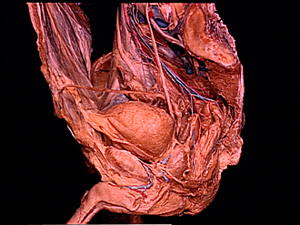
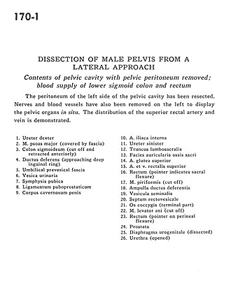
Dissection of male pelvis from a lateral approach
Contents of pelvic cavity with pelvic peritoneum removed; blood supply of lower sigmoid colon and rectum
Image #170-1
KEYWORDS: Large intestine.
Creative Commons
Stanford holds the copyright to the David L. Bassett anatomical images and has assigned Creative Commons license Attribution-Share Alike 4.0 International to all of the images.
For additional information regarding use and permissions, please contact the Medical History Center.
Dissection of male pelvis from a lateral approach
Contents of pelvic cavity with pelvic peritoneum removed; blood supply of lower sigmoid colon and rectum
The peritoneum of the left side of the pelvic cavity has been resected. Nerves and blood vessels have also been removed on the left to display the pelvic organs in situ. The distribution of the superior rectal artery and vein is demonstrated.
- Ureter right
- Psoas major muscle (covered by fascia)
- Sigmoid colon (cut off and retracted anteriorly)
- Ductus deferens (approaching deep inguinal ring)
- Umbilical prevesical fascia
- Urinary bladder
- Pubic symphysis
- Puboprostatic ligament
- Corpus cavernosum of penis
- Internal iliac artery
- Ureter left
- Lumbosacral trunk
- Articular surface of sacrum
- Superior gluteal artery
- Superior rectal artery and vein
- Rectum (pointer indicates sacral flexure)
- Piriform muscle (cut off)
- Ampulla of ductus deferens
- Seminal vesicle
- Rectovesical septum
- Coccyx (terminal part)
- Levator ani muscle (cut off)
- Rectum (pointer on perineal flexure)
- Prostate
- Urogenital diaphragm (dissected)
- Urethra (opened)


