Bassett Collection of Stereoscopic Images of Human Anatomy
Dissection of male pelvis from a lateral approach
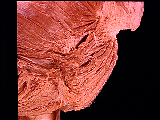
Sphincter ani externus, close-up lateral view
Image #169-3
KEYWORDS: Muscles and tendons.
Creative Commons
Stanford holds the copyright to the David L. Bassett anatomical images and has assigned Creative Commons license Attribution-Share Alike 4.0 International to all of the images.

For additional information regarding use and permissions, please contact the Medical History Center.


Dissection of male pelvis from a lateral approach
Sphincter ani externus, close-up lateral view
The divisions of the sphincter ani externus are not as distinct in this specimen as they are in the preparation shown in view 159-6. The relation of parts of the levator ani muscle to the sphincter ani externus is illustrated here and is also shown in 171-4 after the wall of the anal canal has been opened.
- Iliococcygeus muscle
- Pubococcygeus muscle
- Puborectalis muscle (1-3 are parts of the levator ani muscle)
- Superior fascia of urogenital diaphragm
- Upper pointer: Internal pudendal artery (cutoff) Lower pointer: Pudendal nerve (cut off)
- Urethra (opened)
- Bulb of penis (dissected)
- Bulbospongiosus muscle (cut along midline)
- Coccyx (terminal segment)
- Perineal branch of sacral nerve IV
- Anococcygeal ligament
- Branch of inferior rectal nerve (cut off)
- Area of insertion of Levator ani muscle into wall of anal canal
- Deep part
- Superficial part
- Subcutaneous part (14-16 refer to the external anal sphincter muscle)