Bassett Collection of Stereoscopic Images of Human Anatomy
Male external genitalia and perineum
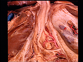
Deep perineal space; bulbourethral gland and duct in situ
Image #168-4
KEYWORDS:
Creative Commons
Stanford holds the copyright to the David L. Bassett anatomical images and has assigned Creative Commons license Attribution-Share Alike 4.0 International to all of the images.

For additional information regarding use and permissions, please contact the Medical History Center.


Male external genitalia and perineum
Deep perineal space; bulbourethral gland and duct in situ
The contents of the deep perineal space or pouch have been dissected. A thin layer of muscle that lay superficial to the bulbourethral gland (9) has been cut away and the gland has been freed of connective tissue.
- Corpus cavernosum of penis left
- Corpus cavernosum of penis right
- Corpus spongiosum of penis
- Bulbospongiosus muscle
- Ramus of ischium
- Upper pointer: Crus of penis Lower pointer: Ischiocavernosus muscle
- Bulb of penis (sectioned in midline)
- Central tendon of perineum
- Bulbourethral gland
- Superficial transverse perineal muscle
- Deep transverse perineal muscle
- Posterior margin of urogenital diaphragm
- Upper pointer: Arcuate pubic ligament Lower pointer: Dorsal nerve of the penis
- Upper pointer: Dorsal artery of penis Lower pointer: Deep artery of penis
- Upper pointer: Spongy part of urethra Middle pointer: Duct of bulbourethral gland Lower pointer: Sphincter muscle of urethra
- Perineal artery (pointer at branching of artery of bulb of penis)