Bassett Collection of Stereoscopic Images of Human Anatomy
Male external genitalia and perineum
Suspensory ligament of penis, left lateral view
Image #167-5
KEYWORDS:
Creative Commons
Stanford holds the copyright to the David L. Bassett anatomical images and has assigned Creative Commons license Attribution-Share Alike 4.0 International to all of the images.

For additional information regarding use and permissions, please contact the Medical History Center.
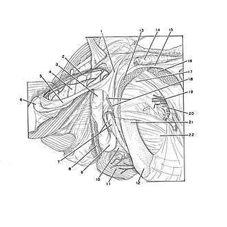
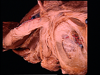
Male external genitalia and perineum
Suspensory ligament of penis, left lateral view
The specimen shown in the preceding view has been turned to expose the left side of the penis. The suspensory ligament (1) is continuous inferiorly with the deep fascia of the penis. It also receives parts of the insertions of the ischiocavernosus and bulbospongiosus muscles (18). The thick tunica albuginea of the left crus of the penis has been removed to reveal the erectile tissue of the crus.
- Suspensory ligament of the penis
- Dorsal nerve of the penis
- Corpus spongiosum of penis
- Corpus cavernosum of penis (covered by tunica albuginea)
- Deep (Buck's) fascia of penis
- Superficial penile fascia
- Upper pointer: Cut edge of tunica albuginea Lower pointer: Erectile tissue of crus of penis
- Bulbospongiosus muscle
- Bulb of penis
- Perineal membrane
- External anal sphincter muscle
- Ramus of ischium
- Tendon of origin of adductor longus muscle
- Spermatic cord (cut across)
- Inguinal ligament
- Pectineus muscle
- Body of pubic bone (periosteum intact)
- Upper pointer: Insertion of bulbospongiosus muscle Lower pointer: Insertion of Ischiocavernosus muscle
- Obturator nerve (emerging from obturator canal)
- Obturator artery
- Inferior pubic ramus
- Obturator membrane