Bassett Collection of Stereoscopic Images of Human Anatomy
Male external genitalia and perineum
Deep perineal fascia
Image #166-4
KEYWORDS: Peripheral nervous system, Vasculature.
Creative Commons
Stanford holds the copyright to the David L. Bassett anatomical images and has assigned Creative Commons license Attribution-Share Alike 4.0 International to all of the images.

For additional information regarding use and permissions, please contact the Medical History Center.
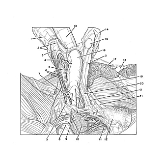

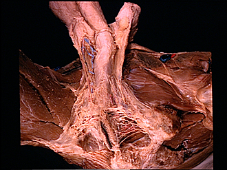
Male external genitalia and perineum
Deep perineal fascia
The superficial perineal fascia (5) has been reflected more completely from the perineal region to expose the deep perineal fascia (Buck's fascia) which encloses the superficial perineal space. The posterior scrotal vessels and nerves have been exposed on the right side and have now been removed from the left side. The superficial and deep fascial layers of the penis (13, 16) may be seen in relation to the fascia of the scrotum and perineum.
- Dartos fascia (enclosing right testis)
- Line of incision separating two halves of scrotum
- Septum of scrotum
- Posterior scrotal vein
- Superficial perineal fascia (membranous layer or Colles' fascia)
- Posterior scrotal nerves
- Bulbospongiosus muscle (faintly visible through deep perineal fascia)
- Ischiorectal fossa
- Central tendon of perineum
- External anal sphincter muscle
- Inferior rectal nerve
- Ischial tuberosity
- Superficial penile fascia
- Dartos fascia enclosing left testis
- Fatty lobule
- Deep (Buck's) fascia of penis (continuous with deep perineal fascia)
- Pectineus muscle (cut across)
- Femoral vein
- Obturator externus muscle
- Ischiocavernosus muscle (covered by deep perineal fascia)
- Ramus of ischium