Bassett Collection of Stereoscopic Images of Human Anatomy
Male external genitalia and perineum
Epididymis dissected, posterior view
Image #165-7
KEYWORDS:
Creative Commons
Stanford holds the copyright to the David L. Bassett anatomical images and has assigned Creative Commons license Attribution-Share Alike 4.0 International to all of the images.

For additional information regarding use and permissions, please contact the Medical History Center.
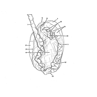
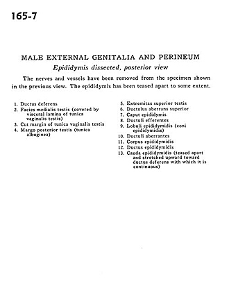
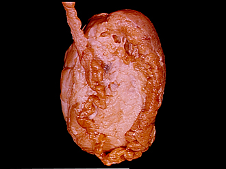
Male external genitalia and perineum
Epididymis dissected, posterior view
The nerves and vessels have been removed from the specimen shown in the previous view. The epididymis has been teased apart to some extent.
- Ductus deferens
- Medial surface of testis (covered by visceral lamina of tunica vaginalis testis)
- Cut margin of tunica vaginalis testis
- Posterior margin testis (tunica albuginea)
- Superior extremity of testis
- Superior aberrant ductule
- Head of epididymis
- Efferent ductules
- Lobules of epididymis (coni epididymidis)
- Aberrant ductules
- Body of epididymis
- Epididymal duct
- Tail of epididymis (teased apart and stretched upward toward ductus deferens with which it is continuous)