Bassett Collection of Stereoscopic Images of Human Anatomy
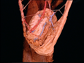
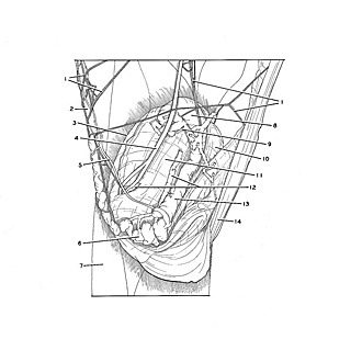
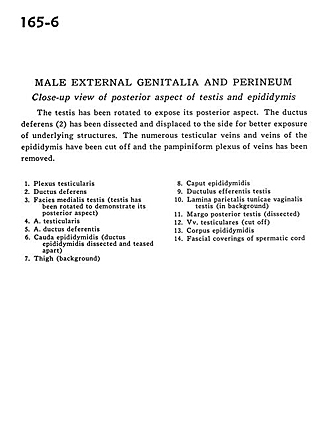
Male external genitalia and perineum
Close-up view of posterior aspect of testis and epididymis
Image #165-6
KEYWORDS:
Creative Commons
Stanford holds the copyright to the David L. Bassett anatomical images and has assigned Creative Commons license Attribution-Share Alike 4.0 International to all of the images.

For additional information regarding use and permissions, please contact the Medical History Center.
Male external genitalia and perineum
Close-up view of posterior aspect of testis and epididymis
The testis has been rotated to expose its posterior aspect. The ductus deferens (2) has been dissected and displaced to the side for better exposure of underlying structures. The numerous testicular veins and veins of the epididymis have been cut off and the pampiniform plexus of veins has been removed.
- Testicular plexus
- Ductus deferens
- Medial surface of testis (testis has been rotated to demonstrate its posterior aspect)
- Testicular artery
- artery of ductus deferens
- Tail of epididymis (ductus epididymidis dissected and teased apart)
- Thigh (background)
- Head of epididymis
- Efferent ductule of testis
- Parietal layer tunicae vaginalis testis (in background)
- Posterior margin testis (dissected)
- Testicular veins (cut off)
- Body of epididymis
- Fascial coverings of spermatic cord