Bassett Collection of Stereoscopic Images of Human Anatomy
Male external genitalia and perineum
Testis and epididymis, close-up anterior view
Image #165-3
KEYWORDS:
Creative Commons
Stanford holds the copyright to the David L. Bassett anatomical images and has assigned Creative Commons license Attribution-Share Alike 4.0 International to all of the images.

For additional information regarding use and permissions, please contact the Medical History Center.
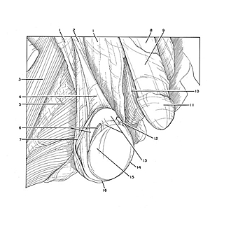
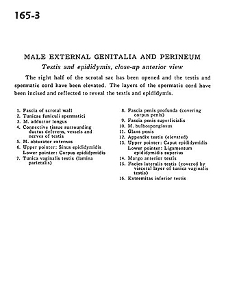
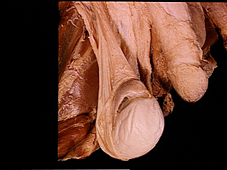
Male external genitalia and perineum
Testis and epididymis, close-up anterior view
The right half of the scrotal sac has been opened and the testis and spermatic cord have been elevated. The layers of the spermatic cord have been incised and reflected to reveal the testis and epididymis.
- Fascia of scrotal wall
- External spermatic fascia
- Adductor longus muscle
- Connective tissue surrounding ductus deferens, vessels and nerves of testis
- Obturator externus muscle
- Upper pointer: Sinus of epididymidis Lower pointer: Body of epididymis
- Tunica vaginalis testis (lamina parietalis)
- Deep (Buck's) fascia of penis (covering body of penis)
- Superficial penile fascia
- Bulbospongiosus muscle
- Glans penis
- Appendix testis (elevated)
- Upper pointer: Head of epididymis Lower pointer: Superior ligament of the epididymidis
- Anterior margin of testis
- Lateral surface of testis (covered by visceral layer of tunica vaginalis testis)
- Inferior extremity of testis