Bassett Collection of Stereoscopic Images of Human Anatomy
Embryo, placenta and fetal membranes
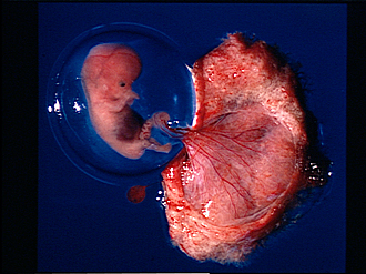
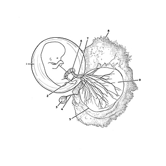
Two-month-old embryo within amniotic sac
Image #164-5
KEYWORDS: Peripheral nervous system.
Creative Commons
Stanford holds the copyright to the David L. Bassett anatomical images and has assigned Creative Commons license Attribution-Share Alike 4.0 International to all of the images.

For additional information regarding use and permissions, please contact the Medical History Center.
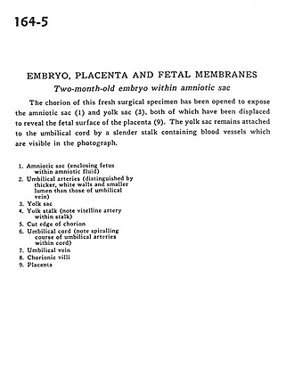
Embryo, placenta and fetal membranes
Two-month-old embryo within amniotic sac
The chorion of this fresh surgical specimen has been opened to expose the amniotic sac (1) and yolk sac (3), both of which have been displaced to reveal the fetal surface of the placenta (9). The yolk sac remains attached to the umbilical cord by a slender stalk containing blood vessels which are visible in the photograph.
- Amniotic sac (enclosing fetus within amniotic fluid)
- Umbilical arteries (distinguished by thicker, white walls and smaller lumen than those of umbilical vein)
- Yolk sac
- Yolk stalk (note vitelline artery within stalk)
- Cut edge of chorion
- Umbilical cord (note spiraling course of umbilical arteries within cord)
- Umbilical vein
- Chorionic VIII
- Placenta