Bassett Collection of Stereoscopic Images of Human Anatomy
Uterus and adnexae
Uterus, uterine tubes and ovaries, anterior aspect
Image #164-2
KEYWORDS: Ovary, Uterus, Muscles and tendons.
Creative Commons
Stanford holds the copyright to the David L. Bassett anatomical images and has assigned Creative Commons license Attribution-Share Alike 4.0 International to all of the images.

For additional information regarding use and permissions, please contact the Medical History Center.
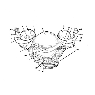
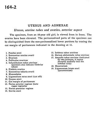
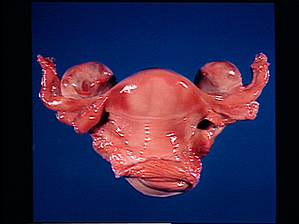
Uterus and adnexae
Uterus, uterine tubes and ovaries, anterior aspect
The specimen, from an 18-year old girl, is viewed from in front. The ovaries have been elevated. The peritonealized parts of the specimen can be distinguished from the non-peritonealized lower portions by tracing the cut margin of peritoneum indicated in the drawing at 11.
- Fundus of uterus
- Uterine extremity of ovary
- Ovary
- Ovarian follicle
- Infundibulum of uterine tube (pointers also indicate tubal fimbriae)
- Ovarian fimbria
- Tubal extremity of ovary
- Mesosalpinx
- Ligamentum teres (of uterus) (cut off)
- Body of uterus
- Cut margin of peritoneum
- Anterior wall of vagina (note vaginal rugae)
- Posterior wall of vagina
- Cervix of uterus
- Isthmus of uterine tube
- Abdominal opening of uterine tube
- Ampulla of uterine tube (indicated by two pointers, it tapers abruptly medially into the isthmus (15)
- Mesovarium
- Broad ligament of uterus (parametrium)