Bassett Collection of Stereoscopic Images of Human Anatomy
Dissection of female pelvis from a lateral approach
Interior of left side of pelvic cavity; sacral plexus; pelvic diaphragm
Image #163-5
KEYWORDS: Muscles and tendons, Peripheral nervous system, Vasculature, Bones joints cartilage.
Creative Commons
Stanford holds the copyright to the David L. Bassett anatomical images and has assigned Creative Commons license Attribution-Share Alike 4.0 International to all of the images.

For additional information regarding use and permissions, please contact the Medical History Center.
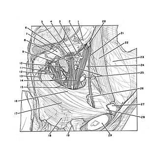
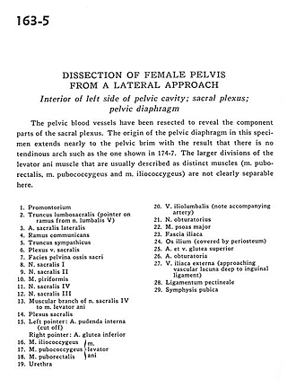
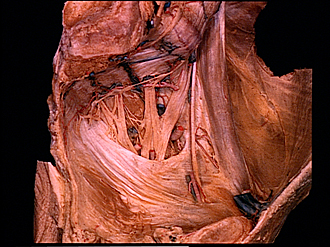
Dissection of female pelvis from a lateral approach
Interior of left side of pelvic cavity; sacral plexus; pelvic diaphragm
The pelvic blood vessels have been resected to reveal the component parts of the sacral plexus. The origin of the pelvic diaphragm in this specimen extends nearly to the pelvic brim with the result that there is no tendinous arch such as the one shown in 174-7. The larger divisions of the levator ani muscle that are usually described as distinct muscles (m. puborectalis, m. pubococcygeus and m. iliococcygeus) are not clearly separable here.
- Promontory
- Lumbosacral trunk (pointer on branch from lumbar nerve V)
- Lateral sacral artery
- Ramus communicans
- Sympathetic trunk
- Sacral venous plexus
- Pelvic surface of sacrum
- Sacral nerve I
- Sacral nerve II
- Piriform muscle
- Sacral nerve IV
- Sacral nerve III
- Muscular branch of sacral nerve IV to levator ani muscle
- Sacral plexus
- Left pointer: Internal pudendal artery (cut off) Right pointer: Inferior gluteal artery
- Iliococcygeus muscle
- Pubococcygeus muscle
- Puborectalis muscle (16-18 comprise the levator ani muscle)
- Urethra
- Iliolumbar vein (note accompanying artery)
- Obturator nerve
- Psoas major muscle
- Iliac fascia
- Ilium (covered by periosteum)
- Superior gluteal artery and vein
- Obturator artery
- External iliac vein (approaching vascular lacuna deep to inguinal ligament)
- Pectineal ligament
- Pubic symphysis