Bassett Collection of Stereoscopic Images of Human Anatomy
Dissection of female pelvis from a lateral approach
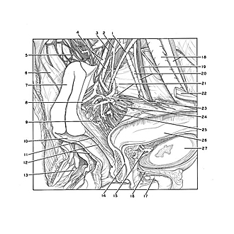
Blood vessels and nerves of uterus, vagina and bladder, medial view
Image #163-2
KEYWORDS: Peripheral nervous system, Uterus, Vagina, Vasculature, Central nervous system, External genitalia, Muscles and tendons, Ovary.
Creative Commons
Stanford holds the copyright to the David L. Bassett anatomical images and has assigned Creative Commons license Attribution-Share Alike 4.0 International to all of the images.
For additional information regarding use and permissions, please contact the Medical History Center.
Dissection of female pelvis from a lateral approach
Blood vessels and nerves of uterus, vagina and bladder, medial view
The uterus and vagina have been retracted posteriorly and the bladder has been drawn forward to expose the vessels and nerves as they approach the left borders of these organs.
- Ureter:
- Uterine artery (note vaginal artery branching off slightly below level of pointer)
- Superior hypogastric plexus
- Uterine veins
- Inferior hypogastric plexus (pelvic plexus)
- Parietal pelvic fascia
- Uterus
- Uterine venous plexus
- Vaginal venous plexus
- Vagina
- Vesicovaginal septum (cut edge)
- Wall of vagina (cut in median plane)
- Anal canal
- Urethra
- Vesical venous plexus
- Dorsal vein of clitoris
- Crus of clitoris
- External iliac artery and vein
- Obturator nerve
- Obturator artery and vein
- Upper pointer: Pelvic diaphragm Lower pointer: Pelvic ganglion
- Ligamentum teres (of uterus) (cut across)
- Superior vesical artery (branch of uterine artery)
- Ureter (at junction with wall of bladder)
- Vesical nerve plexus
- Urinary bladder
- Pubic symphysis


