
Bassett Collection of Stereoscopic Images of Human Anatomy
Dissection of female pelvis from a lateral approach
Nerves and vessels related to pelvic surface of sacrum
Image #162-7
KEYWORDS: Peripheral nervous system, Vagina, Vasculature, Central nervous system, Muscles and tendons.
Creative Commons
Stanford holds the copyright to the David L. Bassett anatomical images and has assigned Creative Commons license Attribution-Share Alike 4.0 International to all of the images.

For additional information regarding use and permissions, please contact the Medical History Center.

Dissection of female pelvis from a lateral approach
Nerves and vessels related to pelvic surface of sacrum
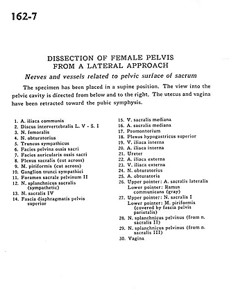
The specimen has been placed in a supine position. The view into the pelvic cavity is directed from below and to the right. The uterus and vagina have been retracted toward the pubic symphysis.
- Common iliac artery
- Intervertebral disc L. V - S. I
- Femoral nerve
- Obturator nerve
- Sympathetic trunk
- Pelvic surface of sacrum
- Articular surface of sacrum
- Sacral plexus (cut across)
- Piriform muscle (cut across)
- Ganglion of sympathetic trunk
- Anterior (pelvic) sacral foramen II
- Sacral splanchnic nerve (sympathetic)
- Sacral nerve IV
- Superior fascia of pelvic diaphragm
- Middle sacral vein
- Middle sacral artery
- Promontory
- Superior hypogastric plexus
- Internal iliac vein
- Internal iliac artery
- Ureter
- External iliac artery
- External iliac vein
- Obturator nerve
- Obturator artery
- Upper pointer: Lateral sacral artery Lower pointer: Ramus communicans (gray)
- Upper pointer: Sacral nerve I Lower pointer: Piriform muscle (covered by parietal pelvic fascia)
- Pelvic splanchnic nerve (from sacral nerve II)
- Pelvic splanchnic nerve (from sacral nerve III)
- Vagina