Bassett Collection of Stereoscopic Images of Human Anatomy
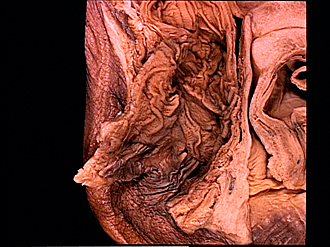
Dissection of female pelvis from a lateral approach
Interior of anal canal and lower part of rectum, close-up view
Image #162-1
KEYWORDS: Anal canal, Large intestine.
Creative Commons
Stanford holds the copyright to the David L. Bassett anatomical images and has assigned Creative Commons license Attribution-Share Alike 4.0 International to all of the images.

For additional information regarding use and permissions, please contact the Medical History Center.


Dissection of female pelvis from a lateral approach
Interior of anal canal and lower part of rectum, close-up view
The lower part of the dissection shown in the preceding photograph is shown in more detail in this close-up view centered on the anal canal.
- Levator ani muscle
- Mucosal tunic of rectum (note numerous small depressions or pits in mucosa marking the sites of lymphatic nodules (folliculi Iymphatici))
- Anal columns
- Pectinate line
- Anal verge
- Hemorrhoidal zone
- Internal anal sphincter muscle
- External anal sphincter muscle
- Plicae transversales of rectum
- Opening of uterus
- Anterior labium
- Posterior wall of vagina
- Vagina
- Upper pointer: Anterior wall of vagina Lower pointer: Vesicovaginal septum
- Muscular tunic of urinary bladder
- Urinary bladder
- Vesical sphincter muscle
- Posterior vaginal wall posterior
- Hymenal caruncles
- Vestibule of vagina
- Levator ani muscle (puborectalis muscle, inserting into wall of anal canal)
- Central tendon of perineum