Bassett Collection of Stereoscopic Images of Human Anatomy
Dissection of female pelvis from a lateral approach
Rectum viewed from right side
Image #161-6
KEYWORDS: Anal canal, Large intestine, Muscles and tendons.
Creative Commons
Stanford holds the copyright to the David L. Bassett anatomical images and has assigned Creative Commons license Attribution-Share Alike 4.0 International to all of the images.
For additional information regarding use and permissions, please contact the Medical History Center.
Dissection of female pelvis from a lateral approach
Rectum viewed from right side
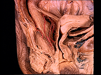
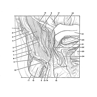
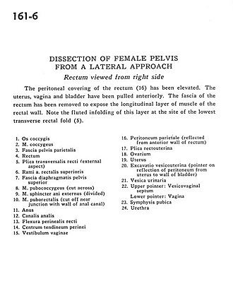
The peritoneal covering of the rectum (16) has been elevated. The uterus, vagina and bladder have been pulled anteriorly. The fascia of the rectum has been removed to expose the longitudinal layer of muscle of the rectal wall. Note the fluted infolding of this layer at the site of the lowest transverse rectal fold (5).
- Coccyx
- Coccygeus muscle
- Parietal pelvic fascia
- Rectum
- Transverse fold of rectum (external aspect)
- Superior branches rectal artery
- Superior fascia of pelvic diaphragm
- Pubococcygeus muscle (cut across)
- External anal sphincter muscle (divided)
- Puborectalis muscle (cut off near junction with wall of anal canal)
- Anus
- Anal canal
- Perineal flexure of rectum
- Central tendon of perineum
- Vestibule of vagina
- Parietal peritoneum (reflected from anterior wall of rectum)
- Rectouterine fold
- Ovary
- Uterus
- Uterovesical pouch (pointer on reflection of peritoneum from uterus to wall of bladder)
- Urinary bladder
- Upper pointer: Vesicovaginal septum Lower pointer: Vagina
- Pubic symphysis
- Urethra


