
Bassett Collection of Stereoscopic Images of Human Anatomy
Dissection of female pelvis from a lateral approach
Rectum, vagina, bladder and ureter, right lateral view
Image #160-7
KEYWORDS: Anal canal, Vagina, Large intestine, Muscles and tendons, Ovary.
Creative Commons
Stanford holds the copyright to the David L. Bassett anatomical images and has assigned Creative Commons license Attribution-Share Alike 4.0 International to all of the images.

For additional information regarding use and permissions, please contact the Medical History Center.

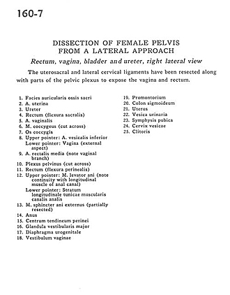
Dissection of female pelvis from a lateral approach
Rectum, vagina, bladder and ureter, right lateral view
The uterosacral and lateral cervical ligaments have been resected along with parts of the pelvic plexus to expose the vagina and rectum.
- Articular surface of sacrum
- Uterine artery
- Ureter
- Rectum (sacral flexure)
- Vaginal artery
- Coccygeus muscle (cut across)
- Coccyx
- Upper pointer: Inferior vesical artery Lower pointer: Vagina (external aspect)
- Middle rectal artery (note vaginal branch)
- Pelvic plexus (cut across)
- Rectum (perineal flexure)
- Upper pointer: Levator ani muscle (note continuity with longitudinal muscle of anal canal) Lower pointer: Longitudinal layer of muscular tunic of anal canal
- External anal sphincter muscle (partially resected)
- Anus
- Central tendon of perineum
- Major vestibular gland
- Urogenital diaphragm
- Vestibule of vagina
- Promontory
- Sigmoid colon
- Uterus
- Urinary bladder
- Pubic symphysis
- Cervix of bladder
- Clitoris