
Bassett Collection of Stereoscopic Images of Human Anatomy
Dissection of female pelvis from a lateral approach
Fascial layers related to bladder, close-up view
Image #160-1
KEYWORDS: Urinary tract, Vasculature.
Creative Commons
Stanford holds the copyright to the David L. Bassett anatomical images and has assigned Creative Commons license Attribution-Share Alike 4.0 International to all of the images.

For additional information regarding use and permissions, please contact the Medical History Center.

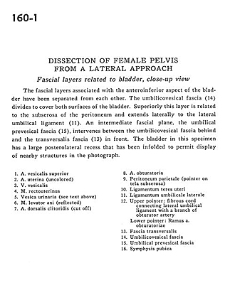
Dissection of female pelvis from a lateral approach
Fascial layers related to bladder, close-up view
The fascial layers associated with the anteroinferior aspect of the bladder have been separated from each other. The umbilicovesical fascia (14) divides to cover both surfaces of the bladder. Superiorly this layer is related to the subserosa of the peritoneum and extends laterally to the lateral umbilical ligament (11). An intermediate fascial plane, the umbilical prevesical fascia (15), intervenes between the umbilicovesical fascia behind the transversalis fascia (13) in front. The bladder in this specimen has a large posterolateral recess that has been infolded to permit display of nearby structures in the photograph.
- Superior vesical artery
- Uterine artery (uncolored)
- Vesical vein
- Rectouterinus muscle
- Urinary bladder (see text above)
- Levator ani muscle (reflected)
- Dorsal artery of clitoris (cut off)
- Obturator artery
- Parietal peritoneum (pointer on tela subserosa)
- Ligamentum teres (of uterus)
- Lateral umbilical ligament
- Upper pointer: Fibrous cord connecting lateral umbilical ligament with a branch of obturator artery Lower pointer: Branch of obturator artery
- Transversalis fascia
- Umbilicovesical fascia
- Umbilical prevesical fascia
- Pubic symphysis