Bassett Collection of Stereoscopic Images of Human Anatomy
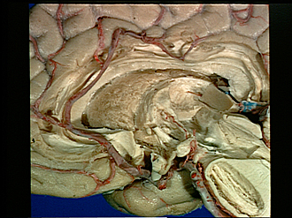
Exploration of the brain from the medial aspect
Caudate nucleus and stria terminalis; thalamic nuclei; medial lemniscus
Image #16-6
KEYWORDS: Brain, Diencephalon, Midbrain.
Creative Commons
Stanford holds the copyright to the David L. Bassett anatomical images and has assigned Creative Commons license Attribution-Share Alike 4.0 International to all of the images.

For additional information regarding use and permissions, please contact the Medical History Center.

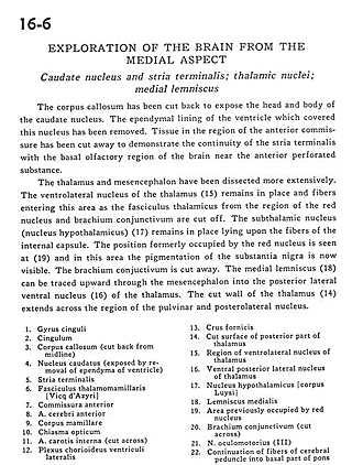
Exploration of the brain from the medial aspect
Caudate nucleus and stria terminalis; thalamic nuclei; medial lemniscus
The corpus callosum has been cut back to expose the head and body of the caudate nucleus. The ependymal lining of the ventricle which covered this nucleus has been removed. Tissue in the region of the anterior commissure has been cut away to demonstrate the continuity of the stria terminalis with the basal olfactory region of the brain near the anterior perforated substance.
- Cingulate gyrus
- Cingulum
- Corpus callosum (cut back from midline)
- Caudate nucleus (exposed by removal of ependyma of ventricle)
- Stria terminalis
- Mamillothalamic tract
- Anterior commissure
- Anterior cerebral artery
- Mamillary body
- Optic chiasm
- Internal carotid artery (cut across)
- Choroid plexus lateral ventricle
- Fornix (ems)
- Cut surface of posterior part of thalamus
- Region of ventrolateral nucleus of thalamus
- Ventral posterior lateral nucleus of thalamus
- Nucleus hypothalamicus
- Medial lemniscus
- Area previously occupied by red nucleus
- Brachium conjunctivum (superior cerebellar peduncle) (cut across)
- Oculomotor nerve (III)
- Continuation of fibers of cerebral peduncle into basal part of pons