
Bassett Collection of Stereoscopic Images of Human Anatomy
Exploration of the brain from the medial aspect
Lateral ventricle; internal cerebral vein within transverse fissure
Image #16-3
KEYWORDS: Brain, Diencephalon, Telencephalon, Vasculature, Ventricules.
Creative Commons
Stanford holds the copyright to the David L. Bassett anatomical images and has assigned Creative Commons license Attribution-Share Alike 4.0 International to all of the images.

For additional information regarding use and permissions, please contact the Medical History Center.
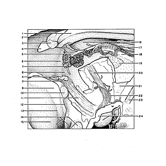
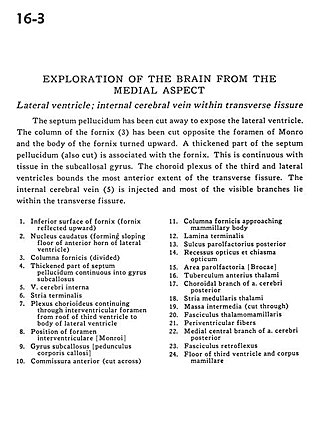
Exploration of the brain from the medial aspect
Lateral ventricle; internal cerebral vein within transverse fissure
The septum pellucidum has been cut away to expose the lateral ventricle. The column of the fornix (3) has been cut opposite the foramen of Monro and the body of the fornix turned upward. A thickened part of the septum pellucidum (also cut) is associated with the fornix. This is continuous with tissue in the subcallosal gyrus. The choroid plexus of the third and lateral ventricles bounds the most anterior extent of the transverse fissure. The internal cerebral vein (5) is injected and most of the visible branches lie within the transverse fissure.
- Inferior surface of fornix (fornix reflected upward)
- Caudate nucleus (forming sloping floor of anterior horn of lateral ventricle)
- Fornix column (divided)
- Thickened part of septum pellucidum continuous into subcallosal gyrus
- Internal cerebral vein
- Stria terminalis
- Choroid plexus continuing through interventricular foramen from roof of third ventricle to body of lateral ventricle
- Position of interventricular foramen (of Monro)
- Subcallosal gyrus
- Anterior commissure (cut across)
- Fornix (column) approaching mammillary body
- Lamina terminalis
- Posterior parolfactory sulcus
- Optic recess and optic chiasm
- Parolfactory area
- Anterior tubercle of thalamus
- Choroidal branch of posterior cerebral artery
- Stria medullaris thalami
- Massa intermedia (cut through)
- Mamillothalamic tract
- Periventricular fibers
- Medial central branch of posterior cerebral artery
- Fasciculus retroflexus
- Floor of third ventricle and mamillary body