Bassett Collection of Stereoscopic Images of Human Anatomy
Exploration of the brain from the medial aspect
Column of fornix, mammillothalamic tract and fasciculus retroflexus
Image #16-2
KEYWORDS: Brain, Diencephalon, Telencephalon.
Creative Commons
Stanford holds the copyright to the David L. Bassett anatomical images and has assigned Creative Commons license Attribution-Share Alike 4.0 International to all of the images.

For additional information regarding use and permissions, please contact the Medical History Center.

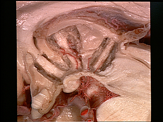
Exploration of the brain from the medial aspect
Column of fornix, mammillothalamic tract and fasciculus retroflexus
Dissection of the medial wall of the thalamus and hypothalamus has now exposed the entire column of the fornix, the mammillothalamic tract and the fasciculus retroflexus. Some gray matter partially covers the latter tract. The internal course of several of the posterior perforating arteries can be followed in the dissected area. Within the meninges filling the transverse fissure a choroidal branch of the posterior cerebral artery is visible.
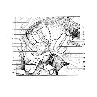
- Fornix (body)
- Choroid plexus third ventricle (note the continuation of this plexus through the interventricular foramen)
- Interventricular foramen (of Monro)
- Mamillothalamic tract
- Anterior commissure (cut across)
- Lamina terminalis
- Subcallosal gyrus
- Tectal part of column of formix
- Mamillary body
- Optic recess
- Infundibular recess and tuber cinereum
- Optic chiasm
- Posterior communicating artery
- Stria medullaris thalami
- Pineal body
- Habenula
- Posterior commissure (cut across)
- Quadrigeminal plate
- Opening of cerebral aqueduct
- Cerebral aqueduct
- Fasciculus retroflexus
- Filaments of oculomotor nerve within tegmentum
- Substantia nigra
- Oculomotor nerve (III)
- Posterior recess of interpeduncular fossa
- Posterior cerebral artery right