
Bassett Collection of Stereoscopic Images of Human Anatomy
Kidneys, suprarenal glands and posterior abdominal vessels, nerves and muscles
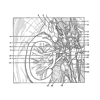
Right kidney and suprarenal gland dissected, close-up view
Image #150-4
KEYWORDS: Adrenal gland, Kidney, Muscles and tendons, Peripheral nervous system, Vasculature.
Creative Commons
Stanford holds the copyright to the David L. Bassett anatomical images and has assigned Creative Commons license Attribution-Share Alike 4.0 International to all of the images.

For additional information regarding use and permissions, please contact the Medical History Center.

Kidneys, suprarenal glands and posterior abdominal vessels, nerves and muscles
Right kidney and suprarenal gland dissected, close-up view
Dissection of the renal parenchyma has exposed the latex-filled arteries and veins within the kidney. Smaller vessels have been trimmed away. The suprarenal cortex has been removed near the center of the gland to expose the brownish medullary tissue as well as to demonstrate the tributaries of the right suprarenal vein within the medulla.
- Right suprarenal vein (cut off at point of emergence from hilus)
- Medulla of suprarenal gland
- Cortex of suprarenal gland
- Right kidney
- Margin of dissected area
- Inferior suprarenal artery (branch of renal artery)
- Filament of renal plexus
- Renal vein (cut off at hilus of kidney)
- Right pointer: Anterior branch of renal artery Left pointer: Interlobar artery
- Renal sinus
- Crus of diaphragm
- Right inferior phrenic artery
- Suprarenal plexus (entering medial margin)
- Celiac trunk
- Celiac ganglion
- Celiac plexus (divided and retracted to expose origin of superior mesenteric artery)
- Superior mesenteric artery
- Middle suprarenal artery (branch of aorta)
- Renal arteries
- Aorticorenal ganglion
- Left renal vein
- Lumbar lymph nodes (lateral aortic nodes)
- Abdominal aorta
- Aortic plexus
- Anterior longitudinal ligament
- Psoas major muscle (covered by fascia)
- Ureter