
Bassett Collection of Stereoscopic Images of Human Anatomy
Kidneys, suprarenal glands and posterior abdominal vessels, nerves and muscles
Right suprarenal gland with inferior vena cava in situ, close-up view
Image #150-2
KEYWORDS: Adrenal gland, Kidney, Muscles and tendons, Peripheral nervous system, Vasculature.
Creative Commons
Stanford holds the copyright to the David L. Bassett anatomical images and has assigned Creative Commons license Attribution-Share Alike 4.0 International to all of the images.

For additional information regarding use and permissions, please contact the Medical History Center.
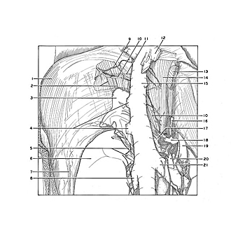
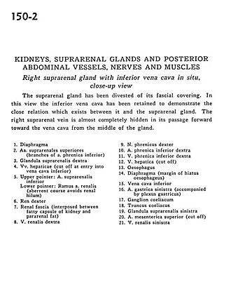
Kidneys, suprarenal glands and posterior abdominal vessels, nerves and muscles
Right suprarenal gland with inferior vena cava in situ, close-up view
The suprarenal gland has been divested of its fascial covering. In this view the inferior vena cava has been retained to demonstrate the close relation which exists between it and the suprarenal gland. The right suprarenal vein is almost completely hidden in its passage forward toward the vena cava from the middle of the gland.
- Diaphragm
- Superior suprarenal arteries (branches of inferior phrenic artery)
- Right suprarenal gland
- Hepatic veins (cut off at entry into inferior vena cava)
- Upper pointer: Inferior suprarenal artery Lower pointer: Branch of renal artery (aberrant course avoids renal hilum)
- Right kidney
- Renal fascia (interposed between fatty capsule of kidney and pararenal fat)
- Right renal vein
- Right phrenic nerve
- Right inferior phrenic artery
- Right inferior phrenic vein
- Hepatic vein (cut off)
- Esophagus
- Diaphragm (margin of esophageal hiatus)
- Inferior vena cava
- Left gastric artery (accompanied by gastric plexus)
- Celiac ganglion
- Celiac trunk
- Left suprarenal gland
- Superior mesenteric artery (cut off)
- Left renal vein