Bassett Collection of Stereoscopic Images of Human Anatomy
Kidneys, suprarenal glands and posterior abdominal vessels, nerves and muscles
Intravenous urogram
Image #150-1
KEYWORDS: Adrenal gland, Kidney, Muscles and tendons, Peripheral nervous system, Vasculature.
Creative Commons
Stanford holds the copyright to the David L. Bassett anatomical images and has assigned Creative Commons license Attribution-Share Alike 4.0 International to all of the images.

For additional information regarding use and permissions, please contact the Medical History Center.

Kidneys, suprarenal glands and posterior abdominal vessels, nerves and muscles
Intravenous urogram
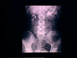
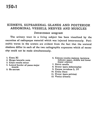
The urinary tract in a living subject has been visualized by the excretion of radiopaque material which was injected intravenously. The peristalticc waves in the ureters are evident from the fact that the ureteral shadows differ in each of the two radiographic exposures which of necessity could not be made simultaneously.
- Rib XI
- Lateral margin of kidney
- Minor renal calyx
- Lateral border of psoas major muscle
- Sacrum
- Major renal calyces (pointers indicate upper, middle and lower major calyces)
- Renal pelvis
- Ureter (abdominal part)
- Body of vertebra L. IV
- Iliac crest
- Ureter (pelvic part)
- Urinary bladder