
Bassett Collection of Stereoscopic Images of Human Anatomy
Exploration of the brain from its lateral aspect
Interventricular foramen, column of fornix and mammillothalamic tract
Image #15-4
KEYWORDS: Brain, Diencephalon, Telencephalon.
Creative Commons
Stanford holds the copyright to the David L. Bassett anatomical images and has assigned Creative Commons license Attribution-Share Alike 4.0 International to all of the images.

For additional information regarding use and permissions, please contact the Medical History Center.

Exploration of the brain from its lateral aspect
Interventricular foramen, column of fornix and mammillothalamic tract
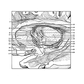
The thalamus and internal capsule have been cut away to expose structures in the vicinity of the interventricular foramen (3). The choroid plexus seen in the central part of the lateral ventricle continues through the interventricular foramen into the roof of the third ventricle.
- Radiation of corpus callosum (cut across)
- Anterior horn lateral ventricle
- Interventricular foramen (of Monro)
- Fornix (column)
- Radiation of rostral lamina of corpus callosum beneath the head of the caudate nucleus
- Posterior part of anterior commissure
- Inner aspect of area parolfactoria
- Optic tract (cut across)
- Limen insulae (cut across lateral to anterior perforated substance)
- Middle cerebral artery
- Amygdaloid nucleus (cut through)
- Central part lateral ventricle
- Choroid plexus lateral ventricle
- Mamillothalamic tract entering anterior nucleus of thalamus
- Stria medullaris thalami
- Thalamus (cut across)
- Periventricular fibers
- Fimbria of hippocampus
- Internal capsule (cut across)
- Hippocampus
- Choroidal branch of posterior cerebral artery
- Choroidal artery (anterior)
- Anterior boundary of inferior horn lateral ventricle