Bassett Collection of Stereoscopic Images of Human Anatomy
Kidneys, suprarenal glands and posterior abdominal vessels, nerves and muscles
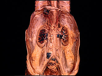
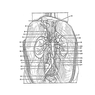
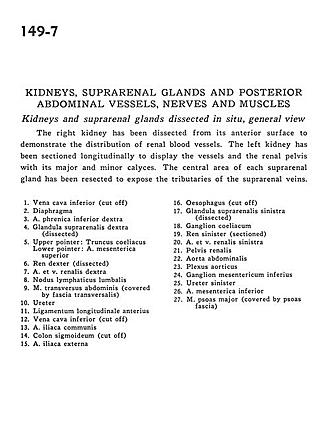
Kidneys and suprarenal glands dissected in situ, general view
Image #149-7
KEYWORDS: Adrenal gland, Kidney, Muscles and tendons, Peripheral nervous system, Vasculature, Overview.
Creative Commons
Stanford holds the copyright to the David L. Bassett anatomical images and has assigned Creative Commons license Attribution-Share Alike 4.0 International to all of the images.

For additional information regarding use and permissions, please contact the Medical History Center.
Kidneys, suprarenal glands and posterior abdominal vessels, nerves and muscles
Kidneys and suprarenal glands dissected in situ, general view
The right kidney has been dissected from its anterior surface to demonstrate the distribution of renal blood vessels. The left kidney has been sectioned longitudinally to display the vessels and the renal pelvis with its major and minor calyces. The central area of each suprarenal gland has been resected to expose the tributaries of the suprarenal veins.
- Inferior vena cava (cut off)
- Diaphragm
- Right inferior phrenic artery
- Right suprarenal gland (dissected)
- Upper pointer: Celiac trunk Lower pointer: Superior mesenteric artery
- Right kidney (dissected)
- Right renal artery and vein
- Lumbar lymph node
- Transversus abdominis muscle (covered by transversalis fascia)
- Ureter
- Anterior longitudinal ligament
- Inferior vena cava (cut off)
- Common iliac artery
- Sigmoid colon (cut off)
- External iliac artery
- Esophagus (cut off)
- Left suprarenal gland (dissected)
- Celiac ganglion
- Left kidney (sectioned)
- Left renal artery and vein
- Renal pelvis
- Abdominal aorta
- Aortic plexus
- Inferior mesenteric ganglion
- Left ureter
- Inferior mesenteric artery
- Psoas major muscle (covered by psoas fascia)