Bassett Collection of Stereoscopic Images of Human Anatomy
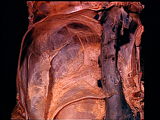
Kidneys, suprarenal, glands and posterior abdominal vessels, nerves and muscles
Right suprarenal gland in situ
Image #148-7
KEYWORDS: Adrenal gland, Kidney, Muscles and tendons, Peripheral nervous system, Vasculature, Overview.
Creative Commons
Stanford holds the copyright to the David L. Bassett anatomical images and has assigned Creative Commons license Attribution-Share Alike 4.0 International to all of the images.

For additional information regarding use and permissions, please contact the Medical History Center.
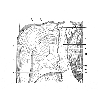

Kidneys, suprarenal, glands and posterior abdominal vessels, nerves and muscles
Right suprarenal gland in situ
A close-up view of the upper area of the specimen shown in the preceding view displays the relation of the right suprarenal gland (10) to the inferior vena cava. The renal fascia (5) continues upward to enclose the gland.
- Diaphragm
- Thoracic cavity
- Right triangular ligament
- Peritoneum and subserous connective tissue (reflected anteriorly)
- Upper pointer: Anterior layer of renal fascia Lower pointer: Right kidney
- Hepatic veins
- Esophagus (emerging below esophageal hiatus)
- Inferior vena cava (small hepatic tributaries cut off along course of vein)
- Crus of diaphragm
- Suprarenal gland (within fascia)
- Suprarenal plexus
- Celiac trunk
- Celiac plexus