
Bassett Collection of Stereoscopic Images of Human Anatomy
Exploration of liver, gall bladder, pancreas, duodenum and spleen
Dissection of duodenum; interior of pars descendens; duodenal papillae
Image #147-3
KEYWORDS: Gallbladder, Liver, Pancreas, Spleen.
Creative Commons
Stanford holds the copyright to the David L. Bassett anatomical images and has assigned Creative Commons license Attribution-Share Alike 4.0 International to all of the images.

For additional information regarding use and permissions, please contact the Medical History Center.
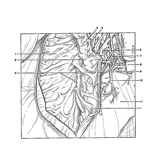
Exploration of liver, gall bladder, pancreas, duodenum and spleen
Dissection of duodenum; interior of pars descendens; duodenal papillae
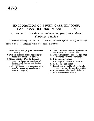
The descending part of the duodenum has been opened along its convex border and its anterior wall has been elevated.
- Plica circularis (in descending part of duodenum)
- Minor duodenal papilla (opening of accessory duct not distinct)
- Upper pointer: Major duodenal papilla (pointer on openings of common bile duct and pancreatic duct) Lower pointer: Longitudinal duodenal fold (forming frenulum of duodenal papilla)
- Muscular layer of duodenum (pointer on cut edge of a circular fold)
- Muscular layer of duodenum (pointer indicates circular fibers)
- Pancreatic duct
- Accessory pancreatic duct
- Superior mesenteric artery
- Prominent hood-like plica circularis overlying major duodenal papilla
- Inferior pancreaticoduodenal artery
- Horizontal part of duodenum