Bassett Collection of Stereoscopic Images of Human Anatomy
Exploration of liver, gall bladder, pancreas, duodenum and spleen
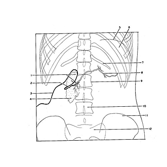
Radiograph of pancreatic ducts
Image #147-2
KEYWORDS: Gallbladder, Liver, Pancreas, Spleen.
Creative Commons
Stanford holds the copyright to the David L. Bassett anatomical images and has assigned Creative Commons license Attribution-Share Alike 4.0 International to all of the images.
For additional information regarding use and permissions, please contact the Medical History Center.
Exploration of liver, gall bladder, pancreas, duodenum and spleen
Radiograph of pancreatic ducts
The radiopaque contrast medium has been injected into the main pancreatic duct (8) in a living subject through a catheter which was passed into the duct at the duodenal papilla. The injection mass also filled the accessory duct (1) and passed out in the duodenum through the opening of this duct. (This film was obtained through the courtesy of Dr. H. Doubilet of New York University.)
- Upper pointer: Contrast medium within duodenum Lower pointer: Accessory pancreatic duct (point at which tributary enters duct from lower part of head of pancreas)
- Approximate location of duodenal papilla (catheter and contrast medium in pancreatic duct difficult to differentiate)
- Catheter
- Duodenum (inferior part, containing some opaque material)
- Diaphragm (radiolucent area above is lung)
- Spleen
- Rib XII
- Pancreatic duct
- Vertebra L. II
- Body of vertebra L. IV
- Ilium
- Sacrum.


